 |
||
|
Digital Diagnostic Imaging Recommendations
Recommendations on the Delivery of, Access to and
The move from an analogue, or screen based x-ray capture to Digital Diagnostic Imaging capture and distribution is inevitable, however many clinicians may not have had sufficient exposure or understanding to appreciate the complex technical specifications involved in such changes. This web site provides a reference and background for those wishing to embrace such information to ensure that the transition to digital imaging facilitates optimal patient care consistent with the local clinician and imaging provider capabilities and resources. The options detailed here specifically address the situation where access to diagnostic quality imaging is required - where the clinician will use their own interpretation of the diagnostic images to guide treatment decisions, rather than rely entirely on the Radiologist's report.
Where non-diagnostic review or educational image access is required, these recommended benchmarks do not necessarily apply. The viewing clinician however must be aware that such images, while useful for review purposes, may not of a sufficient standard to allow optimal diagnostic analysis. Follow the links below to access DDI (Digital Diagnostic Imaging) Options and potential technology suppliers:
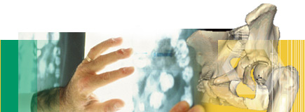
What is a diagnostic image ?A key issue is the definition of what constitutes “diagnostic quality imaging”. It is recognised that imaging requirements will vary according to the clinical situation, the patient’s status and the technique used to capture the images. The ultimate responsibility for assessment of the suitability of images rests with those practitioners involved. This document aims to provide guidance however without the intention of being prescriptive. For the purpose of this document, the definition of a diagnostic quality image is provided by the RANZCR and ADIA: “An image that is comparable in quality and presentation to that used by radiologists in reporting”. The key features, from the clinical perspective, are that such images will be of high resolution and capable of supporting clinical measurement. To be of value to a clinician, the images must also be in a form that is accessible, convenient and appropriate to the clinical circumstances. For practical purposes the discussion about what constitutes a diagnostic quality digital image starts with the implementation of uncompressed DICOM standard format images. DICOM is the international standard for digital images in healthcare.
Images of lesser quality may also be produced which while of great clinical use and helpful for clinical review and patient education, may or may not be determined by the treating practitioner as being suitable for “diagnostic use” in a specific clinical situation.
Imaging can be divided into three (not necessarily mutually exclusive) broad categories:
This web site predominantly addresses category 1, and more specifically, the process and form in which such images are made available to the clinician looking after the patient. It aims to provide guidance to clinicians who have a need to review diagnostic quality images in the course of patient care. A number of specific imaging needs related to certain medical specialties (e.g. cardiology, radiation oncology) are not specifically covered at this stage, however the common utilisation of the DICOM format does mean that the image data can be distributed and accessed using the same principles. It is also recognised that significant technological advances in diagnostic imaging have led to a variety of image formats and functionality requiring innovative solutions to maximize the effectiveness of delivery, access, manipulation and archiving. Variations in display matrices and bit depth, three dimensional display methods, multi planar reconstruction, Image fusion, dynamic imaging and colour rendering all support the progressive move from a film based delivery approach to digital format. However, when patient care decisions depend upon the review of the diagnostic images, those responsible for the ultimate clinical management of the patient must be involved in any decision to adopt a material change in the way images are provided. The transition to digital imaging must not compromise the diagnostic and treatment capabilities of the clinician, and should facilitate, not hinder, appropriate image access at the patient care interface. Similarly, it is important that referrers have realistic expectations in requesting “film” in all cases. Multislice CT studies often create over 1,000 images per patient study, and an element of selection must be applied judiciously when key images are filmed. When selected images are supplied on film (or other hard copy) they must be appropriate to the clinical setting. Equally, it is not always possible to anticipate at the outset all the possible uses of, and requirements for, a diagnostic imaging examination. While precise communication between the referrer and the provider can minimise difficulties concerning examination type, processing and delivery, there should also be a mechanism whereby additional appropriate requirements (e.g. those arising from on-referral) can be met expeditiously.
Data Content of Diagnostic Imaging Examinations In considering contemporary imaging, it is useful to differentiate between images such as plain radiographs (large matrix) and those generated by other modalities, including CT, MRI and ultrasound (small matrix). The diverse matrix sizes, and the variable numbers of images generated by the various modalities, pose different challenges in image display. Plain (digital) radiography typically utilizes large matrix (e.g. 2000 x 3000) resolution images. The recommended minimum resolution for diagnostic quality is 2.5lp/mm. This equates to approximately 127ppi, and at 10 or more bits per pixel, the major challenge is the size of the image and the need for a large high quality monitor to allow full size display, and adequate network or transfer capacity as images range from 12 – 40MB in size. Cross-sectional images obtained from CT and MRI is typically small matrix (e.g. 512 x 512) at 12 - 14 bit images, and is more easily shared digitally. They are captured digitally, and individual image size is generally less than 1MB. The major challenge from these modalities is the large numbers of images often obtained (~ 100 – 3000) and the collective size of the entire data set.
Imaging modalities classed by data content Within Diagnostic Imaging there are three broad groups of image data sets (with some overlap between these): 1. Plain Radiography
2. Dynamic and Complex Diagnostic Imaging
3. Cross-sectional imaging (of a defined volume)
Anatomical Reference Images for Cross-sectional imaging In the delivery of cross-sectional images, either digitally or by hard copy, it is critical that a spatial location guide , indicating the relationship of each cross-sectional image to standard anatomical landmarks, be provided (these may be known as ‘scout images’ or ‘pilot’ views, topograms, etc.) This applies whether the images are delivered on film or on digital media, however for digital media this should be part of the functionality of the DICOM reading software.
Diagnostic-quality images distributed in digital format must comply with DICOM standards and IHE PDI profiles if distributed on portable media. For both small and large-matrix digital image data, the data set should provide full resolution data for processing, manipulation and subsequent display if required depending on the clinical requirements. Compression
|
|||||||
 |
©
2007 - 2026 clinPACS | disclaimer
| legal notice website design by WorldWeb | credits |